Life Processes Notes for Class 10 Science
Life Processes- The basic functions performed by living organisms to maintain their life of this earth are called life processes. The basic life processes common to all the living organisms are: Nutrition and Respiration, Transport and Excretion, Control and Coordination (Response to stimuli), Growth, Movement and Reproduction.
Energy is needed for the life Processes and food is a kind of fuel that provides energy to all the living organisms.

Nutrition: – It is the method of obtaining nutrients from the environment.
Nutrition is a process of intake of nutrients (like carbohydrates, fats, proteins, minerals, vitamins and water) by an organism as well as the utilisation of these nutrients by the organism
Modes of Nutrition: – There are mainly two modes of nutrition:
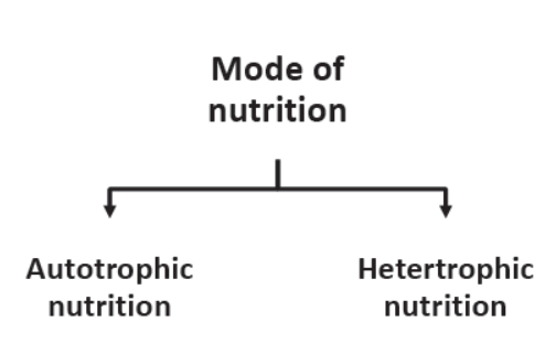
Autotrophic mode of Nutrition: – The organisms which make their own food from carbon dioxide and water in the presence of sunlight and chlorophyll are called autotrophs. These organisms are also called producers and include green plants and some bacteria.
Heterotrophic mode of Nutrition: – Those organisms which cannot make their own food from inorganic substances like carbon dioxide and water, and depend on other organisms for their food are called heterotrophs. Ex.
Animals (man, dog, cat etc.) & yeast.
Types of Heterotrophic Nutrition: – Can obtain its food from other organisms in three ways. So the heterotrophic mode of nutrition is of three types:

Saprotrophic Nutrition: – Saprotrophic nutrition is that nutrition in which an organism obtains its food for decaying organic matter of dead plants, dead animals and rotten bread etc. ex. Fungi (like bread moulds, mushrooms, yeast).
Parasitic Nutrition: – The parasitic nutrition is that nutrition in which an organism derives its food from the body of another living organism (called its host) without killing it. Ex Plants like Cuscuta (amarbel) and some animals like Plasmodium and roundworms.
Holozoic Nutrition: – The holozoic nutrition is that nutrition in which an organism takes the complex organic food materials into its body by the process of ingestion; the ingested food is digested and then absorbed into the body cells of the organism. Ex man, cat, dog, deer, tiger, lion, bear, giraffe, frog, fish and Amoeba etc.
Nutrition in Plants: – Green plants are autotrophic and synthesize their own food by the process of photosynthesis.
Photosynthesis: – The process by which green plants make their own food (like glucose) from carbon dioxide and water by using sunlight energy in the presence of chlorophyll is called photosynthesis.
The process of photosynthesis can be represented as:

The process of photosynthesis takes place in the green leaves of a plant.
In plants and most algae, it occurs in the chloroplasts and there two principal reactions:
1. Light reactions (light-independent) bring about the photolysis of water.

2. Dark reaction (light-independent) during this reaction carbon dioxide is reduced to carbohydrate in a metabolic pathway known as the Calvin cycle.
Events occurring during photosynthesis process.
1. Absorption of light energy by chlorophyll.
2. Conversion of light energy to chemical energy and splitting of water molecules into hydrogen and oxygen.
3. Reduction of carbon dioxide to carbohydrates.
Chloroplast: Any of the chlorophyll containing organelles (i.e. plastid) which are found in large numbers in plant and algae cells undergoing photosynthesis is called chloroplast. The plant chloroplasts are typically lens-shaped and bounded by a double membrane.
Site of Photosynthesis in Plants: Chloroplasts are the main site of photosynthesis and occur in the mesophyll cells of the leaf.
Raw materials for Photosynthesis: Carbon dioxide, water, chlorophyll and sunlight are the essential raw materials for photosynthesis.
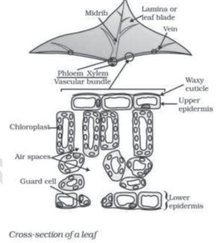
1. Carbon dioxide is a gas, which is released into the atmosphere during respiration.
2. Water is another requirement for photosynthesis. Which is transported upward through xylem tissues to the leaves, from where it reaches the photosynthetic cells?
3. Chlorophyll it is a green pigment in plants which acts as a catalyst. It is responsible for absorption of the sun’s energy by the plant. The chlorophyll pigments are photoreceptor molecules which play a key role in the photosynthetic process.
4. Light affects photosynthesis by its intensity quality and duration. In green light, the rate of photosynthesis is minimum, while in red and blue lights the rate of photosynthesis is maximum.
Stomata: They are the tiny pores present on the epidermal surface of the leaves.
The function of stomata is gas exchange between the plant and the atmosphere.
Each stoma is bordered by two semicircular kidney shaped guard cells.

Opening and Closing of Stomatal pore. The opening and closing of the pore is a function of the guard cells.
The guard cells swell when water flows into them causing the Stomatal pore to open. Similarly the pore closes if the guard cells shrink.
As large amount of water is lost through these stomata, the plant closes these pores when it does not require carbon dioxide for photosynthesis.
Heterotrophic Nutrition: The type of nutrition in which organisms derive their food (nutrients) from living organisms. In heterotrophic nutrition the energy is derived from the intake and digestion of the organic substances, normally of plant or animal tissue.
Nutrition in Amoeba: The mode of nutrition in amoeba is holozoic and it is omnivorous.


- It feeds on unicellular plant or animal such as Paramoecium, Oscillatoria, etc.
- he various steps of nutrition are ingestion, digestion, assimilation and egestion.
- hen Amoeba comes in contact with food particles, it sends out pseudopodia which engulf the prey by forming a food-cup. This process is ingestion.
- hen the tips of the encircling pseudopodia touch each other, the food is encaptured into a bag called food vacuole. This step is digestion.
- he food vacuole serves as a temporary stomach secretion digestive juice.
- he digested food gets absorbed and diffuses into the cytoplasm and then assimilated.
- gestion of undigested food takes place at any point on the surface of the body; i. e. there is no fixed anus.
Human digestive system: The organs which are responsible for ingestion, digestion, absorption, assimilation and egestion constitute the digestive system. The digestive system comprises of the alimentary canal and associated digestive glands.

A. Alimentary canal in man is 9 meters long and consists of the following parts:
1. Mouth. It leads into buccal cavity. The floor of the buccal cavity has a tongue bearing taste buds (sweet, salt, sour & bitter). Man possesses teeth on both the jaws. There are 32 teeth of four different types, namely incisors, canines, premolars and molars.
2. Pharynx. It is a short, conical region that lies after the mouth cavity. The pharynx is divided into two parts the nasopharynx which lies behind the nasal cavities and the oropharynx which lies behind the mouth.
3. Oesophagus. It is a long, narrow, muscular tube which leads to the stomach.
4. Stomach. It lies below the diaphragm on the left side of abdominal cavity and is J-shaped. The food is stored and partly digested in the stomach.
5. Small Intestine. It is a convoluted tube and differentiated into three regions, viz., duodenum, which is the first part of small intestine and is curved C-shaped; jejunum, comparatively longer and more coiled, and ileum, which is the last part of small intestine whose inner surface is folded to form villi, which absorbs the products of digestion.
6. Large Intestine. It is much shorter and wider than small intestine and is differentiated into three regions viz., caecum, which is small rounded blind sac from which vermiform appendix arises; colon is the inverted U-shaped tube and the rectum opens to exterior through anus.
B. Digestive Glands. Various glands associated with alimentary canal are:
1. Salivary Glands. The salivary glands secrete the first of the digestive juices, the saliva. There are three pairs of salivary glands namely the parotids (largest salivary gland, lie on sides of face) sub-maxillary (lie at angles of lower jaw) and sublingual glands (under front part of teeth).

2. Gastric Glands. They are branched tubular glands which lie in the mucus membranes of the stomach. They secrete gastric juice, which are clear, acidic containing HCI, enzymes and mucus.
3. Liver. It is the largest gland in man and lies below diaphragm in the right upper part of abdomen. Liver comprises or two lobes right and left, where the right lobe is much larger than the left lobe. The cells of liver, i.e., hepatic cells produce bile juice which flows out of liver through hepatic ducts forming common bile duct and opens into duodenum. Bile juice then flows into gall bladder through the cystic ducts.

4. Pancreas. It is a soft lobulated gland present in between the loops of duodenum. It secrets pancreatic juice containing enzymes which is poured into duodenum with the help of pancreatic duct.

Note: Dental Caries: It is the tooth decay which involves destruction of the enamel layer of the tooth by acids produced by the action of bacteria on sugar. If dental caries is not treated, it can spread to the dentine and pulp of the tooth, causing inflammation and infection of the tooth.
Process of Nutrition: The process of nutrition involves five steps:
1. Ingestion. It is the process of taking food inside the body. The food taken by man is masticated by the teeth before swallowing.
2. Digestion. It is the process of conversion of large, complex and insoluble organic molecules into simpler, smaller and soluble molecules.
- Digestion may be intracellular (Paramoecium) or extracellular (multicellular animals).
- The process of digestion starts in the mouth cavity and continues upto the intestine.
- In the mouth, food gets mixed up with saliva secreted by salivary glands.
- Saliva contains an enzyme salivary amylase which breaks polysaccharide starch into disaccharide maltose.
The food from the mouth cavity passes into the stomach through the oesophagus.
The gastric glands of the stomach secrete gastric juice which contains hydrochloric acid, two protein digesting enzymes—pepsin and renin, mucus and small amount of gastric lipase.
- Pepsin breaks down proteins into peptones in acidic medium of gastric juice.
- Muscles present on the wall of stomach churn and propel the food forward.
- The digested food moves from stomach to duodenum of the small intestine.
- Duodenum receives juices from liver, i.e., bile and pancreatic juice from pancreas.
- The pancreatic juice contains trypsin, amylase and lipase.
- The proteins, fats and carbohydrates are further digested into amino acids, glycerol, fatty acids, glucose and fructose.
- Finally, the digestion is completed in the ileum with the secretion of the intestinal juice by intestinal glands.
- The intestinal juice consists of amylolytic, proteolytic and lipolytic enzymes.
3. Absorption. It is the process of mixing of digested food in the body fluid. All the digested food is absorbed in the ileum. The food is absorbed by diffusion, osmosis or by active participation of the cells of the intestine.
4. Assimilation. It is the process of utilisation of absorbed food for various body functions. The absorbed nutrients are utilized to resynthesize complex molecules like carbohydrates, proteins and fats inside the cells.
5. Egestion. It is the process of elimination of undigested food formed in the cells, or in the lumen of large intestine (colon and rectum) through the anus.
Table: Summary of the digestive enzymes of various glands with their secretions and end products of Digestion in Man


Cellular Respiration: It is the process of biochemical oxidation of nutrients in the presence of specific enzymes at optimum temperature in the mitochondria of cells to release energy for various metabolic activities.
Respiration is a catabolic process and there occurs exchange of gases, viz., oxygen and carbon dioxide, between the body and the outside environment. It is of two types: – aerobic and anaerobic respiration.
1. Aerobic Respiration. When tissues carry out oxidation of food materials, utilizing molecular oxygen; the process is called aerobic respiration.

2. Anaerobic Respiration. When cells or organisms carry out oxidation of nutrients without utilizing molecular oxygen; the process is called anaerobic respiration.

Breakdown of Glucose by Various Pathways:

ATP. It refers to a nitrogenous compound. Adenosine Triphosphate. The energy released during cellular respiration is immediately used to synthesis a molecule called ATP from ADP and inorganic phosphate as

ATP is used to fuel all activities in the cell. Therefore, it is said to be the energy currency for most cellular processes.
Respiration in Plants: It takes place in all parts of a plant-like root, stem and leaf
- Exchange of gases in roots take place by the process of diffusion, where oxygen diffuses into the root hairs and passes into the root cells, from where carbon dioxide moves out into the soil.
- In woody plants, bark has lenticels for gaseous exchange.
- In leaves, respiration also takes place by diffusion of oxygen through stomata into cells of the leaf and carbon dioxide is released into the atmosphere, when its concentration in cells increases.
Respiration in Animals: It takes place with the help of some specific respiratory organs which differs in different animal groups, according to their habitat. Aquatic animals like fish, prawns and mussels have gills as respiratory organs; land animals like lizard, bird, human have lungs, frogs breathe both by skin and lungs and insects like grasshopper, hou8sefly or cockroach have air tubes or trachea as their respiratory organs.

Human respiratory system: Lungs are the respiratory organs in humans and are located in the cavity of thorax.
- Human respiratory system consists of nostrils, nasal cavities, pharynx, trachea, bronchi, and bronchioles leading to alveoli inside the lungs.
- This kind of respiration, where lungs are the main structures is called pulmonary respiration.
- Respiratory system communicates with the outside atmosphere through external nostrils which draw air into nasal cavities.
- The nasal cavities open into the internal nostrils through which air enters the pharynx.
- The pharynx leads into trachea or windpipe through a slit called glottis.
- The trachea runs down the neck and enters the thorax and divides into the right and left bronchi.
- These two tubes enter into two elastic and conical lungs; which are enclosed in double-walled sacs called pleura.
- The bronchi within lungs, branch into smaller tubes called bronchioles; and each bronchiole opens into many thin-walled balloon like structure called alveoli.
- The alveoli provide a surface for gaseous exchange.
- The walls of the alveoli contain an extensive network of blood-vessels.

Mechanism of Breathing in Human: Breathing is a complex mechanical process involving muscular movement that alters the volume of the thoracic cavity and thereby that of the lung.
- Breathing occurs involuntarily but its rate is controlled by the respiratory center of the brain.
- The space of thoracic cavity increases or decreases by outward and inward movements of the ribs caused by external intercostal and internal intercostal muscles.
- This action is also assisted by the contraction and expansion of the diaphragm,
- The floor of the thoracic cavity is completely closed by diaphragm. It is a thin muscular septum separating the abdominal and thoracic cavities.
- The inhalation and exhalation of the air take place continuously in the respiratory system.
- Inspiration or inhalation in concerned with the taking in of atmospheric air or oxygen into the thoracic cavity. It is possible only when the volume of the thoracic cavity increases and the pressure of the contained air in the thoracic cavity decrease.
- Expiration or exhalation is concerned with the expelling of carbon dioxide from lungs. It takes place when the volume of the thoracic cavity decreases and the pressure of the contained air in the thoracic cavity increases.
Gas Exchange in Alveoli:-
- Blood rich in carbon dioxide, i.e., the deoxygenated blood enters the capillary network of alveolus.
- CO2 diffuses into the alveolar cavity because of its higher concentration in the blood,
- Alveolus has a higher concentration of oxygen as compared too the blood in capillaries.
- Therefore, O2 diffuses into the capillaries and combines with haemoglobin of red blood cells to form oxyhaemoglobin to be transported throughout the body.
Gas Exchange in Tissues:
- In the cells, continuous metabolism of glucose and other substances results in the production of CO2 and utilisation of O2.
- In the cells and tissues, fluid concentration of oxygen decreases while the concentration of CO2 increases.
- Therefore, oxyhaemoglobin breaks down releasing O2 which diffuses out from the capillaries into the tissue fluid and then into each and every cell.

Transportation in Human Beings: In humans, transportation of oxygen, nutrients, hormones and other substances to the tissues, CO2 to the lungs and waste products to the kidneys us carried out by a well-defined circulatory system.
Circulatory System: It comprises of the heart, blood vessels, blood, lymphatic vessels and lymph, which together serve to transport materials throughout the body.
Blood: It is a bright red-coloured liquid connective tissue that circulates in the entire body by the muscular pumping organ, the heart. The volume of blood is about 6 litres in and adult human body.
Plasma: it is the liquid part of the blood excluding blood cells.
- Plasma consists of water in which many substances are dissolved including plasma proteins (albumin, globulin, fibrinogen and antibodies), salts (sodium and potassium chlorides and bicarbonates), food substances (amino acids, glucose, and fats), hormones, digested and waste excretory products.
- In the plasma, RBCs, WBCs and blood platelets are immersed.
- Plasma without fibrinogen is called serum.
Blood Corpuseles:
1. Red blood Corpuscles (RBCs) or Erythrocytes. These are minute circular biconcave discs having no nucleus.
They look red due to the presence of red coloured pigment, haemoglobin.
2. White Blood Corpuscles (WBCs) or Leucocytes. These are large, nucleated colourless cells and are numerous than erythrocytes, WBCs are larger than RBCs.
WBCs are mainly of two types – Granulocytes and Agranulocytes.
Agranulocytes — 2 Subtypes (a) Monocytes (b) Lymphocytes.
Granulocytes – 3 Subtypes (a) Basophils (b) Eosinophils (c) Neutrophils
3. Blood Platelets. Platelets are rounded, colourless, biconvex and non-nucleated blood cells; which help in coagulation of blood. They are called thrombocytes; they are formed in bone marrow.
Function of Blood: Blood performs the following functions:
1. Transport of Oxygen. Red blood corpuscles contain haemoglobin that combines with oxygen to from oxyhaemoglobin which is transported to the tissues of the tissues of the body for the purpose of respiration.
2. Transport of Carbon dioxide. Carbon dioxide produced by the tissues as a result of respiration is transport is transported by the blood plasma and also by the haemoglobin to the lungs from where it is removed.
3. Transport of Nutrients. The digested and absorbed nutrients like glucose, amino acids, fatty acids, vitamins, etc. are first transported to the liver and then to the whole of tissues for their storage, oxidation and synthesis to new substances.
4. Transport of Excretory Products. Nitrogenous wastes like ammonia, urea and uric acid of body are transported to the kidneys by the blood from where they are eliminated.
5. Regulation of Body Temperature. The blood flows in all parts of the body, so it equalizes the body temperature. It carries heat produced from one place to another place of the body.
6. Maintenance of pH. The plasma proteins act as buffer system and maintain required pH of the body tissues.
7. Transport of Hormones. The plasma of blood transports various hormones from one region to another and brings about the co-ordination in the working of the body.
8. Water Balance. The blood maintains water balance at constant level by distributing uniformly over the body.
9. Protection from Diseases. The WBC (eosinophils, neutrophils, monocytes) engulf the bacteria and other disease causing organisms by phagocytosis. The lymphocytes produce antibodies against the invading antigens.
10. Clotting of Blood. Blood forms a clot at the site of injury, thus preventing further loss of blood. Blood helps in rapid healing of wounds.
Our Pump – The Heart: The heart is a pumping organ that receives blood from the veins and pumps in into the arteries. It is situated in thoracic cavity which lies above the diaphragm between the two lungs. It is enclosed in a double-walled membraneous sac, the pericardium.

A. Chambers of the Heart. The interior of the heart is divided into four chambers which receive the circulatory blood.
1. The Atria (Auricles). The two superior chambers are called the right and left atria. The atrias are separated by apartition called the inter-atrial septum. The sinuatrial node (SAN) or the pacemaker is located in the upper wall of the right atrium.
2. The Ventricles. The two interior chambers of the heart are the right and left ventricles. They are separated from each other by and inter-ventricular septum.
B. Valves of the Heart. Valves are muscular flaps which prevent the blood to flow through it. Two types of heart valves are distinguished:
1. TheAtrioventricular Valves. These valves separate the atria from the ventricles. The right side of the heart possesses the tricuspid valve or right atrio-ventricular valve and the left side of the heart possesses the bicuspid or mitral valve.
2. Semilunar Valves. These are located in the arteries leaving the heart. The pulmonary semilunar valve lies in the opening where the pulmonary trunk leaves the right ventricle and aortic semilunar valve lies at the opening between the left ventricle and aorta.
C. Blood Flow through the Heart:
1. The right atrium receives deoxygenated blood from all parts of the body through large veins called vena cava.
2. When the right atrium is full of blood, it contracts and the tricuspid valves open under pressure and the blood is forced into the right ventricle.
3. When the right ventricle is full of blood, it contracts and the blood is pumped into the pulmonary trunk.
4. The pulmonary trunk divides into the right and left pulmonary each of which carries the blood to the lungs for oxygenation.
5. The oxygenated blood returns to the heart via the pulmonary veins that empty into the left auricle.
6. When the left auricle contracts, the blood passes into the left ventricle by the opening of the bicuspid valve.
7. On contraction of the left ventricle, the blood is pumped into the largest artery called aorta.
8. The aorta branches into vessels which transport blood to the heart and all body parts.
Double Circulation in Man: The circulatory system of man is called double circulation as the blood passes through the heart twice in one complete cycle of the body. It involves two circulations:
1. Pulmonary Circulation. This circulation is maintained by the right side of the heart.
- It begins in the right ventricle which expels the blood into the pulmonary trunk.
- The blood flowing into the vascular system of the lungs becomes oxygenated and returns to the heart (left atrium) through pulmonary veins.

2. Systemic Circulation. This circulation is maintained by the left ventricle which sends the blood into the aorta.
- The aorta divides into arteries, arterioles and finally to capillaries and thereby supplies oxygenated blood to various parts of the body.
- From there, deoxygenated blood is collected by venules which join to form veins and finally, vena cava and pour blood back into the heart.
Heart of different Vertebrates: The separation of right side and left side of the heart is useful to keep away oxygenated and deoxygenated blood fro mixing. This separation allows a highly efficient supply of oxygen to the body which is useful in animals having high energy needs such as birds and mammals. These animals use energy to maintain their body temperature. In animals, like reptiles and amphibians, their body temperature depends on temperature of the environment. They have three chambered hearts and tolerate some mixing of oxygenated and deoxygenated blood streams. Fish have only two chambered heart and blood pumped to gills is oxygenated and passes directly to the rest of the body. Thus, blood goes only once through the heart in fishes during one cycle of passage through the body. But it does through the heart twice during each cycle in other vertebrates, i.e. by double
circulation.
Blood Pressure. It is the force that blood exerts against the wall of a vessel. This pressure is much greater in arteries than in veins.
- The pressure of blood artery during contraction or ventricular systole is called systolic pressure and pressure in artery during relaxation or ventricular diastole is called diastolic pressure.
- The normal systolic pressure is about 120 mm of Hg and diastolic pressure is 80 mm of Hg.
- Blood pressure is measured using an instrument called a sphygmomanometer.
- Abnormally high blood pressure called hypertension can lead to rupture of an artery and internal bleeding.
Blood Vessel. There are three types of blood vessels of different sizes involved in blood circulation, viz., arteries, veins and capillaries, which are all connected to form a continuous closed system.

- Arteries are wide and thick, elastic-walled vessels that carry oxygenated blood from the heart to different organs of the body. Aortais the main artery.
- Veins are thin-walled, having valves that carry deoxygenated blood from different organs to the heart.
- Capillaries. The artery divides into smaller vessels on reaching an organs or tissue which bring blood in contact with all the individual cells. These smallest vessels which have one cell thick wall are called capillaries. These walls are permeable, so that water and dissolved substances pass in and out, exchanging oxygen, carbon dioxide, dissolved nutrients and excretory products with the tissues.
Blood clotting: It is the mechanism that prevents the loss of blood at the site of an injury or wound by forming a blood clot. The blood has platelet cells which circulate around the body and plug these leaks by helping to clot the blood at these points of injury to prevent it from excessive bleeding. The major events in blood clotting or coagulation are given in this flow chart:

Lymphatic System. It is a system of tiny tubes called lymph vessels or lymphatics and lymph nodes or lymph glands in the human body which transport the liquid, lymph from the body tissues to the blood circulatory system.
Lymphatic system runs parallel to veins and consists of the following parts:
- Lymph or tissue fluid is colourless containing lymphocyte cells which fight against infection. Lymph flows only in one direction, i.e., from tissues to heart. Lymph is also called extracellular fluid as it lies outside the cells. Lymph drains into lymphatic capillaries.
- Lymphatic Capillaries are thin-walled capillaries forming a network in every organ except nervous system.
- Lymphatic Vessels from second pathway for fluid returning from the tissues to the heart. The lymphatic capillaries unite to form lymphatic vessels which are very small veins in structure.
- Lymph Nodes or Lymph Glands are situated in the course of the lymph vessels and generally occur in groups and are oval or kidney shaped. They are rich with phagocytes and lymphocytes, thus act as filters for the microorganisms.
Functions of Lymph:
1. Lymph carries digested and absorbed fat from intestine and drains excess fluid from extra cellular space back into the blood.
2. It protects the body by killing the germs and draining it out of the body tissues with the help of lymphocytes contained in the lymph nodes.

Transportation in Plants: Plant transport system moves energy stored from leaves and raw materials from roots.
These two pathways are constructed as conducting tubes—xylem, which moves water, minerals obtained from the soil; and phloem which transports products of photosynthesis from the leaves where they are synthesized to other parts of the plant.
Conducting Tissue. It is a tissue comprising of xylem and phloem that carries substances from one part of the plant body to another. Within the vascular bundle, the xylem is internally located while phloem lies towards the outside of the organ.
Transport of Water and Minerals. Plants require water for making food by photosynthesis and also need mineral salts for leaves and flowers.
Xylem. It is a tissue that transports water and dissolved mineral nutrients from the roots to all other parts of the vascular plant. Xylem consists of four kinds of elements:
I. Vessels (b) Tracheids (c) Xylem fibers (d) Xylem parenchyma
Mechanism of Transport of Water and Minerals in a Plant:
- The vessels and tracheids of roots, stems and leaves in xylem tissue are interconnected to form continuous system of water conducting channels reaching all parts of the plant.
- The cells of the roots in contact with the soil actively take up ions which creates a difference in the ion concentration between the root and the soil.
- Thus, there is steady movement of water into root xylem from the soil, creating a column of water that is pushed upwards.
- Plant uses another strategy to move water in the xylem upwards to the highest points of the plant body.
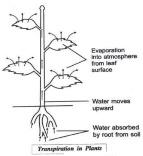
- The water which is lost through the stomata is replaced by water from the xylem vessels in the leaf.
- Evaporation of water molecules from the cells of a leaf creates a suction which pulls water from the xylem cells of roots.
- This loss of water is transpiration which helps in the absorption and upward movement of water and minerals dissolved in it from roots to the leaves.
- Transpiration becomes the major driving force in movement of water in the xylem during the day when the stomata are open.
- This mechanism is also known as cohesion of water theory or transpiration pull.
Importance of Transpiration: –
1. Ascent of Sap. It is the upward movement of cell sap, i.e., water and minerals through the xylem.
2. Removal of Excess Water. Transpiration helps to remove excess water.
3. Cooling Effect. Transpiration kelps to regulate the temperature of the plant – since evaporation reduces temperature.
4. Absorption and Distribution of Salts. The continuous water current produced by transpiration helps to absorb and distribute the salts.
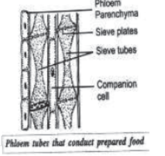
Transport of Food and Other Substances. The food (i.e., sugar and other metabolites synthesized in the leaves and substances like hormones synthesized in the leaves and substances like hormones synthesized at the tips of roots and stems are transported to other parts of the plant through a conducting tissue called phloem.
Phloem. It is a vascular tissue that conducts food materials in vascular plants from regions where they are produced, i.e., leaves to regions such as growing points, where they are needed for the purpose of storage or consumption.
Translocation. It is the transport of food from the leaves to other parts of the plant and occurs in the part of the vascular tissue known as phloem.
Mechanism of Transport of Food and Other Substances in a Plant:
- The translocation of food and other substances takes place in the sieve tubes with the help of adjacent companion cells both in upward and downward directions.
- The translocation in phloem is achieved by utilizing energy from ATP.
- The food entering the phloem tubes in the leaves is transported to all other parts of the plant by the network of phloem tubes present in all parts of the plant like stem and roots.
- Translocation is necessary because every part of the plant needs food for obtaining energy, for building its parts and maintaining its life.
Excretion. It is the biological process of elimination of harmful metabolic waste products from the body of an organism. The mode of excretion is different in different organisms. Many unicellular organisms remove these wastes by simple diffusion from the body surface into the surrounding; while complex multicellular organisms use specialized organs for excretion. The organs that are involved in this process constitute the excretory system.

Excretion in Human Beings. The excretory system of human beings collects and drains out the wastes from the body. It consists of a pair of kidneys, a pair of ureters, a urinary bladder and a urethra.
1. Kidneys. It is the main excretory organ.
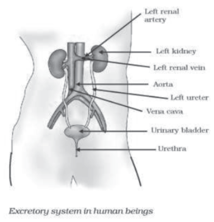
- Each kidney is bean-shaped, reddish brown in colour and is located in the abdomen, one on either side of the backbone.
- The left kidney is placed a little higher than the right kidney.
- The renal artery brings in the uncleaned blood containing waste substances into the kidneys.
- The renal vein carries away the cleansed blood from the kidneys.
2. Ureters or Excretory Tubes. They are the thin muscular tubes coming out from each kidney which opens into the urinary bladder. Ureters are ducts which drain out urine from the
kidneys.
3. Urinary Bladder. It is a pear-shaped reservoir that stores urine before being discharged to the outside.

4. Urethra. It is a muscular tube that arises from the neck of the bladder and conducts the urine to the outside through an opening at its end, the urinary opening.
Functions of the Kidney:
1. It removes the poisonous substances such as urea, other waste salts and excess water from the blood and excretes them in the form of yellowish liquid called urine.
2. It regulates the osmotic pressure/water balance of the blood.
3. It regulates pH of the blood.
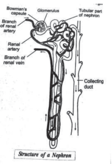
Nephrons. Each kidney is made up of a large number of excretory filtration units called nephrons or uriniferous tubules. These are considered as the functional unit of kidney.
- It consists of a long coiled tubule whose one end is connected to the double walled cup shaped structure of Bowman’s capsule and its other end to a urine-collecting duct of a kidney.
- he Bowman’s capsule contains a bundle of blood capillaries which is called glomerulus.
- The function of glomerulus is to filter the blood passing through it.
- The function of tubular part of nephron is to allow selective reabsorption of the useful substances into the blood capillaries.
Formation of Urine. The purpose of urine is to filter out waste products from the blood.
- The nitrogenous waste such as urea or uric acid are removed from blood in the kidneys, thus kidneys are the basic filtration unit.
- ach capillary cluster in the kidney is associated with the cup-shaped end of a tube that collects the filtered urine.
- Each kidney has large numbers of these filtration units called nephrons.
- Some substances in the initial filtrate such as glucose, amino acids, salts and a major amount of water are selectively reabsorbed as the urine flows along the tube. This depends on how much excess water is there in the body and on how much of dissolved waste is there to be excreted.
- The urine formed in each kidney enters a long tube, the ureter which connects the kidneys with the urinary bladder.
- Urine is stored in the urinary bladder until the pressure of the expanded bladder leads to pass out through the urethra.
Artificial Kidney. It is a device to remove nitrogenous waste products from the blood through dialysis. In case of kidney failure and artificial kidney can be used.
Dialysis. It is the procedure used in artificial kidney to replace a non-functional or damaged kidney. In the process, blood of the patient is allowed to pass through the long cellulose tubes dipped in a tank containing dialysing solution having same ionic concentration as plasma. The waste substances diffuse out of blood into the tank and the clean blood is returned back into the patient through a vein.
Excretion in plants. Plants produce a number of waste products during their life processes.
- The main waste products produced by plants are carbon dioxide, water vapour and oxygen.
- Plants get rid of excess water by transpiration.
- The gaseous wastes of respiration and photosynthesis in plants (carbon dioxide, water vapour and oxygen) are removed through the ‘stomata’ in leaves and ‘lenticels’ in stems and released in the air.
- Many plant waste products are stored in cellular vacuoles. Wastes products may be stored in leaves that fall off, other waste products are stored as resins and gums.
- Plants excrete some waste substances into the soil around them.
- Some of the plant wastes which are useful to humans are—Natural rubber, gum, resins and essential oils like sandalwood oil, eucalyptus oil, clove oil and lavender oil.


